Palghar, Maharashtra
Giemsa Stain
| Business Type | Manufacturer, Supplier |
| C.A.S. No. | 51811-82-6 |
| Solubility 0.1 % (MeOH) | Clear solution |
| Absorption Maximum(in MeOH) ?1max | 640-650 nm |
| Click to view more | |
Preferred Buyer From
| Location | Anywhere in India |
Product Details
Solubility
Insoluble in water (<0.1%), methanol (5.8 mg/l), and ethanol.
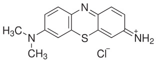
Giemsa Stain is a laboratory reagent and stain for microscopy. A compound of eosin with methylene blue and its oxidation products. When blood films are stained using Giemsa stain, the nucleus and cytoplasm of white blood cells take on a characteristic blue or pink coloration. Giemsa stain functions by rearranging chromatin and inducing G bands producing a high quality stain for chromatin and the nuclear membrane. Giemsa stain is used to differentiate nuclear and/or cytoplasmic morphology of platelets, RBCs, WBCs, and parasites (1,2). The most dependable stain for blood parasites, particularly in thick films, is Giemsa stain containing azure B. Liquid stock is available commercially. The stain must be diluted for use with water buffered to pH 6.8 or 7.0 to 7.2, depending on the specific technique used.
Applications:
Giemsa and May Grünwald solutions are intended for use in staining blood films or bone marrow films. Solutions are for “In Vitro Diagnostic Use.” Giemsa stain is a buffered thiazine-eosinate solution designed to provide coloration of blood cells similar to the original product described by Giemsa. It may be used separately or in combination with a May Grünwald Stain, also available from Sigma-Aldrich. Giemsa stain is a biological stain for thin blood films to differentiate leucocytes, for thick blood films to show malarial parasites, and for bone marrow to show cell morphology. It is also used as a chromosome stain and to differentiate nuclear morphology of platelets, RBCs, and other cell types. Giemsa Stain was designed primarily for the demonstration of parasites in malaria, but it was also employed in histology because of the high-quality staining of the chromatin and the nuclear membrane, the metachromasia of some cellular components, and the different qualities of cytoplasmic staining depending on the cell type. The use of methylene azure and its mixture with methylene blue to form an eosinate made stable the stain and its results. Giemsa's stain is regarded as the world's standard diagnostic technique for malaria's plasmodium, and it is also the basic stain for classifying lymphomas in the Kiel classification.
Looking for "Giemsa Stain" ?
Explore More Products

